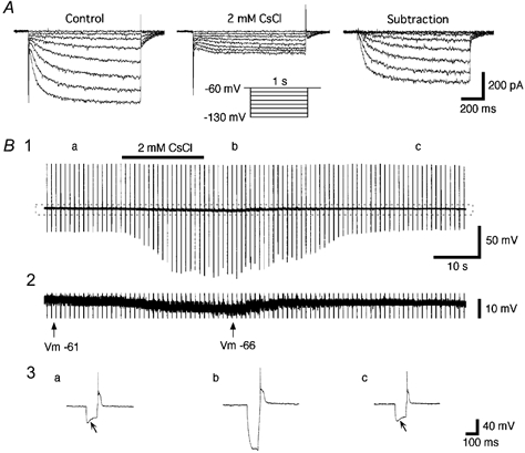
Figure 4. Extracellular Cs+ blocks Ih.

A, currents elicited by hyperpolarizing voltage steps (inset) in control (modified ACSF, left panel) and after addition of extracellular CsCl (2 mM, middle panel). The Cs+-sensitive current was isolated by digital subtraction (right panel). B1, recordings of the membrane potential and input resistances before, during and after the bath application of CsCl (2 mM). The bar above the trace indicates the period of the application of CsCl. The input resistance was measured by injection of hyperpolarizing current pulses (160 pA, 100 ms duration) once every 5 s. Note the marked increases in input resistance induced by the extracellular Cs+. B2, an enlargement of the portion indicated by the dotted rectangle in B1 shows clearly identifiable membrane hyperpolarization (about −5 mV) during exposure to extracellular Cs+. B3, enlargements of the voltage responses to the hyperpolarizing current pulses before, during and after the bath application of Cs+. Traces are from the points indicated by a, b and c in B1. The oblique arrows point to the voltage ‘sag’. Note that the blockade and recovery of the voltage ‘sag’ is coincident with the membrane potential (Vm) changes.
