
Figure 2. Ultrastructure of AIL cells and myocytes.
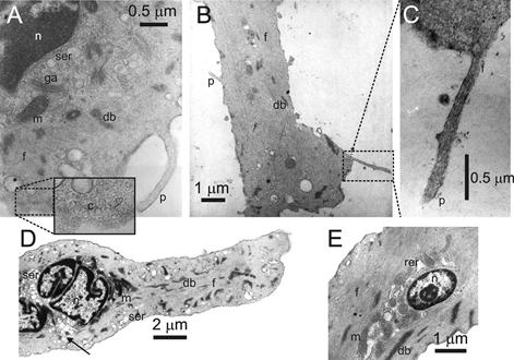
Transmission electron micrographs of AIL cells (A, B and C) and of myocytes (D and E). n, nucleus; ser, smooth endoplasmic reticulum; rer, rough endoplasmic reticulum; ga, golgi apparatus; m, mitochondrion; db, dense body; f, filaments; c, caveolae (inset in A); p, thin process.