
Figure 5. Inward and outward current in voltage-clamped cells.
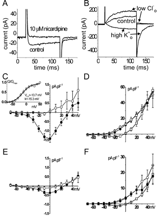
A, original traces of inward current in AIL cells obtained by stepping from the holding potential of −60 mV to 0 mV for 100 ms. I−V relationship for the inward current in AIL cells (C) and myocytes (E). •, control; ○, with nicardipine (10 μm). Inset in C, activation curve of inward current in AIL cells. G/Gmax, relative conductance; V0.5, voltage of half-maximal activation; k, slope factor. The inward current of AIL cells was ≈4 times more dense than in myocytes. Nicardipine abolished the inward current in AIL cells and inhibited it in myocytes. B, original traces of outward current (100 ms steps from −60 mV to −10 mV) in AIL cells. I−V relationship for the outward current in AIL cells (D) and myocytes (F). •, control; ○, in low [Cl−]o physiological salt solution (PSS); □, in high [K+]o PSS. The outward current of AIL cells was ≈twice as dense as in myocytes. In both the AIL cells and myocytes, increasing the [K+]o to 120 mm shifted the curve to the right and lowering the [Cl−]o to 33.4 mm did not change the outward current significantly.