
Figure 10. Localisation of AIL cells in the arteries.
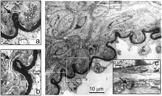
Transmission electron micrograph of a transverse section of mesenteric artery. Rectangles delineated by the broken line show areas which are shown enlarged in a, b and c. l, vessel lumen; ec, endothelial cell; bl:,basal lamina; n, nucleus. Arrows indicate processes. The cells with thin processes can be seen in the media, both in the layer immediately under the basal lamina (a and b) and deeper in the media (c).