
Figure 5. Granular layer stimulation evokes briefer parallel fibre EPSCs than molecular layer stimulation in GLAST knock-out mice.
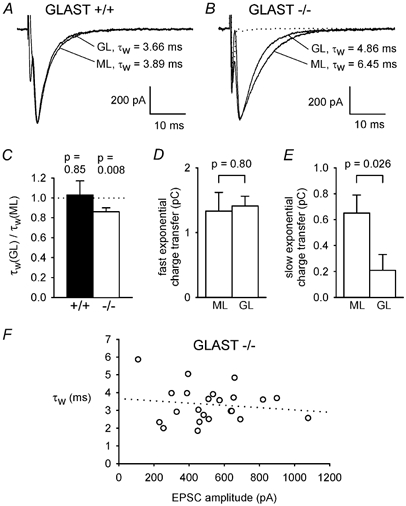
A, specimen parallel fibre EPSCs of similar amplitude recorded in the same wild-type cell (+/+) when stimulating the granule cell axons either in the molecular layer (ML) or the granular layer (GL), with weighted decay time constants (τw) indicated. B, as for A but in a GLAST knock-out cell (−/-). C, ratio of τw for GL and ML stimulation in 6 wild-type and 12 knock-out cells. P values are for t tests comparing data with unity. Amplitudes of EPSCs were 653 ± 72 pA for GL and 660 ± 52 pA for ML stimulation in the +/+ cells, and 578 ± 68 pA for GL and 578 ± 70 pA for ML stimulation in the −/− cells (not significantly different: P = 0.94 for +/+ and 0.998 for −/−). D and E, charge transfer by the faster exponential component (D), and by the slower exponential component of eqn (1) (E), for ML and GL stimulation in the 12 knock-out cells. The amplitudes and time constants of the fast and slow exponential components of the EPSC decay (in the order Afast, τfast, Aslow, τslow) were: for ML stimulation, 467 ± 58 pA, 3.34 ± 0.41 ms, 47.5 ± 18.1 pA, 9.12 ± 1.90 ms (7 out of 12 cells showed a slow component); and for GL stimulation: 484 ± 55 pA, 2.99 ± 0.20 ms, 19.9 ± 14.1 pA, 14.5 ± 2.8 ms (4 out of 12 cells showed a slow component). F, τw as a function of EPSC amplitude for GL stimulation in 23 knock-out cells (includes some cells stimulated only in the GL layer). This experiment (unlike that of Fig. 3E) required inclusion of data from different cells because the range of EPSC amplitudes which can be generated by altering the intensity of GL stimulation is much smaller than for ML stimulation.