
Figure 2. Voltage-gated potassium currents and the effect of TNBS ileitis on current density.
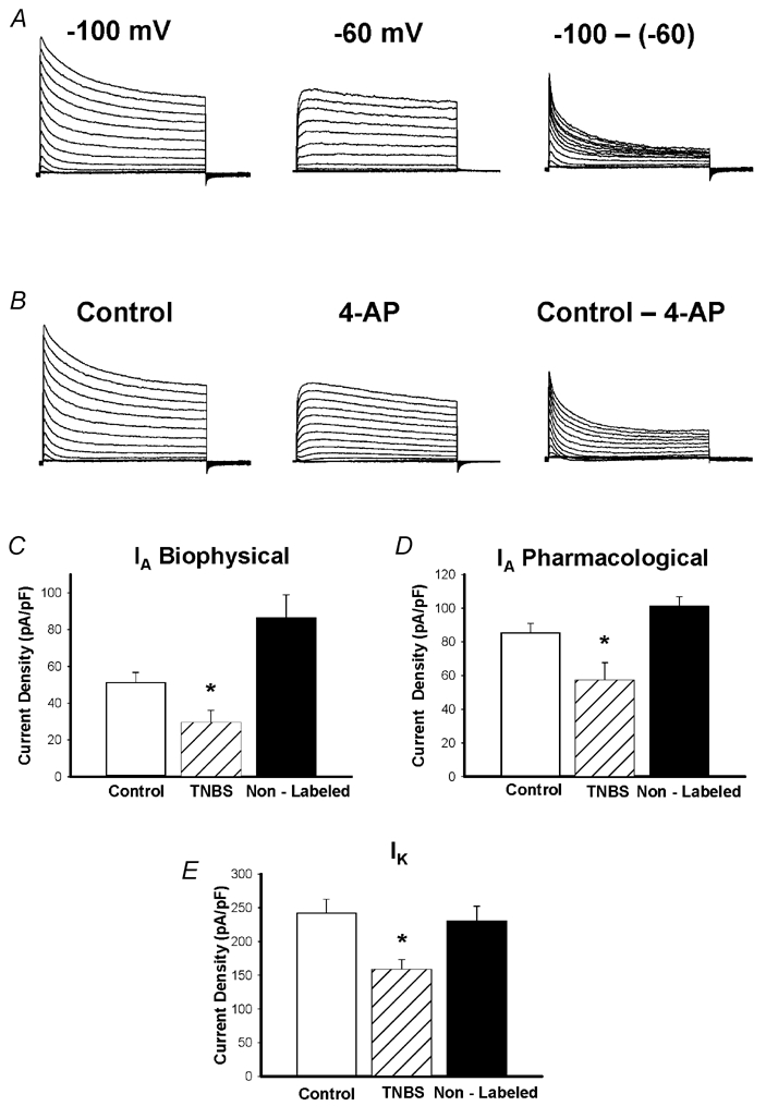
Representative traces from one cell showing currents separated biophysically by manipulating the holding potential (A) and pharmacologically by 4-AP (B). Currents were elicited in response voltage steps from −90 mV to +50 mV in 5 mV increments. A, left: at holding potentials of −100 mV, two currents were apparent, a transient, inactivating ‘A’ type current, and a non-inactivating sustained IK type current. A, middle: resultant current when membrane potential is held at −60 mV. Note disappearance of the transient component. Only the sustained component remains, and was measured at 400 ms. A, right: subtraction of the sustained from the total current yields IA. IA amplitude was measured as the peak of the transient component. B, left: in the pharmacological experiments, the holding potential was −100 mV. B, middle: when the voltage steps were repeated in the presence of 1 mm 4-AP the transient component was significantly inhibited. B, right: results of subtracting current obtained in the presence of 4-AP from control, revealing the 4-AP-sensitive IA current. C, effect of TNBS ileitis on mean peak IA density obtained using either biophysical (left) or pharmacological separation (right). TNBS ileitis results in a significant reduction in IA density compared to control, or non-labelled TNBS neurons (*P < 0.05). D, effect of TNBS ileitis on mean peak IK density. TNBS ileitis results in a significant reduction in peak IK density, compared to control and non-labelled cells (*P < 0.05).