
Figure 2. Amplification of the frequency response to corticospinal tract stimulation (A and B) and muscle stretch (C and D) during increasing firing frequencies evoked by gradually increasing depolarizing current through the recording microelectrode.
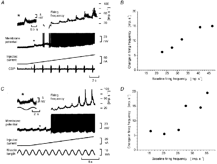
A, recordings from a posterior biceps motoneurone. The inset (*) shows the CST EPSP evoked by a train (50 stimuli at 300 Hz) to the pyramidal tract, recorded at a membrane potential close to firing threshold. The main recordings show the cell's firing frequency (upper plot), spike train (second trace) the injected current (third trace), and the cord dorsum potential (CDP, fourth trace). B, the increased firing frequency during the CST excitation (ordinate) as a function of firing frequency induced by the injected current through the recording microelectrode (abscissa). C, recordings from a triceps surae motoneurone. The inset (*) shows the stretch evoked EPSP recorded at a membrane potential close to firing threshold. The main recordings show the cell's firing frequency (upper plot), spike train (second trace), injected current (third trace), and muscle stretch (fourth trace). D, the increased frequency response during the stretch excitation (ordinate) as a function of firing frequency induced by the current through the recording microelectrode (abscissa).