
Figure 6. Temperature dependence of resting intracellular pH.
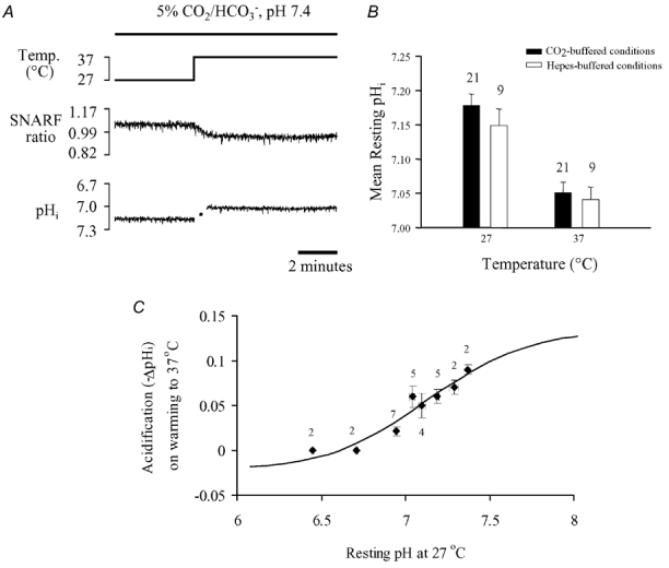
A, effect of raising bath temperature from 27 to 37°C on carboxy-SNARF-1 fluorescence ratio (middle trace) and corresponding pHi (bottom trace) in 5 % CO2/HCO3−-buffered conditions, pHo 7.35–7.40 at 37°C. B, mean resting pHi measured at 27 and 37°C for 5 % CO2/HCO3−-buffered (filled columns) and Hepes-buffered (open columns) conditions. C, pooled data (mean ±s.e.m.) of the change in pHi (-ΔpHi) following an increase in bath temperature from 27 to 37°C in Hepes-buffered conditions. Numbers beside symbols denote the number of observations. Note that for n = 2, the upper and lower s.e.m. values represent the individual data points. Low initial pHi levels were generated by pre-pulsing with 15 mm NH4Cl in the presence of 30 μm Hoe 642 (see text for details). The continuous line shows predictions of the dual-buffer model described in the Appendix.