
Figure 3. Ca2+ speeds up the upstroke of the action potential.
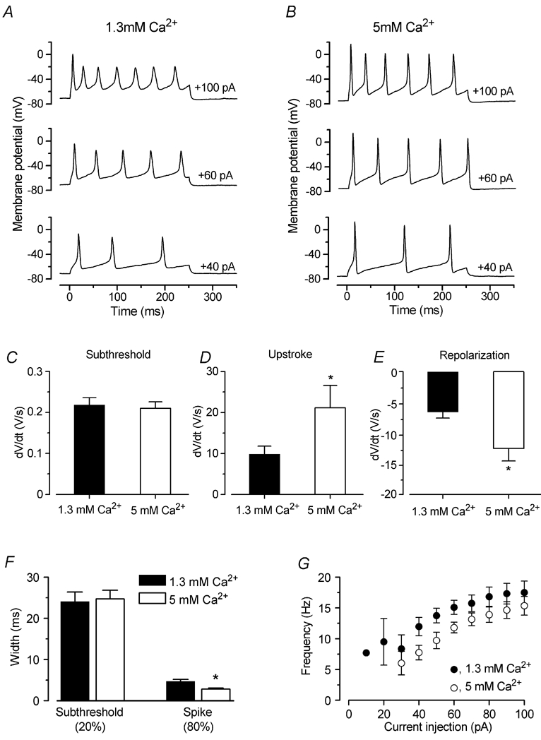
A and B, voltage responses from an IHC before and during superfusion of 1.3 and 5 mm extracellular Ca2+, respectively. Current steps were applied in 10 mV increments up to +100 pA, and for clarity only a few examples are shown. Note that in the presence of 5 mm Ca2+ the action potentials became faster and reached a more depolarized potential. The recording conditions were: P7, Cm 8.4 pF; Rs 5.0 MΩ; temperature 37°C. C-E, rate of the subthreshold depolarization, and the rise and fall of the action potentials, respectively, during superfusion of 1.3 and 5 mm Ca2+ (P7, n = 8). F, widths measured at the subthreshold (20 %) and spike (80 %) level as shown in Fig. 8A (dashed lines). G, frequency of evoked action potentials before and during superfusion of 5 mm Ca2+ as a function of depolarizing current injection (P7, n = 8).