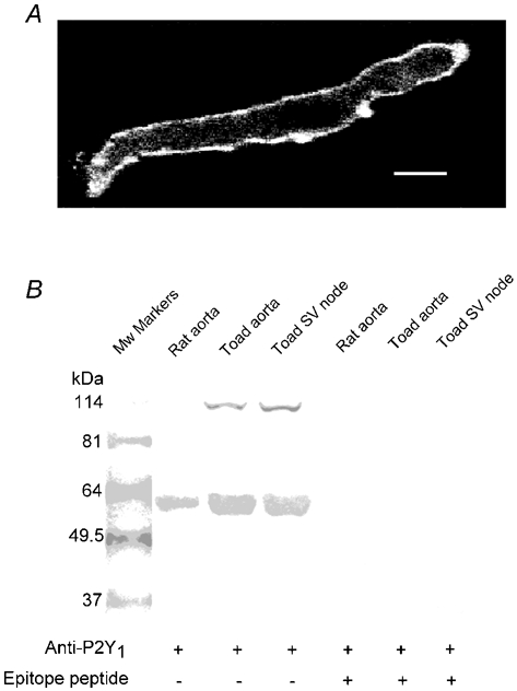
Figure 6. Immunohistochemistry and Western blotting of P2Y1 purinoceptors.

A, pacemaker cell fixed and stained with P2Y1 antibody and fluorescent secondary antibody. Bar indicates 6 μm. B, Western blots demonstrating P2Y1 expression in toad tissues. Total protein extracts were subject to 10 % SDS-PAGE and probed with P2Y1 antiserum in the absence (lanes 2–4) and presence (lanes 5–7) of epitope peptide. One band of about 57 kDa was observed in the rat aorta sample that is known to express P2Y1 receptors. This band is probably the glycosylated form of P2Y1. The same band was present in the toad samples, whereas an additional band of about 114 kDa was also seen in these samples. This band may be a dimer of the P2Y1 receptor. Preincubation with the epitope peptide greatly attenuated the 114 kDa band and abolished the 57 kDa band, suggesting specific labelling.
