
Figure 2. Distribution of rSK1 protein in HEK 293 cells.
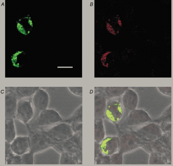
A, confocal image of HEK 293 cells transiently transfected with a YFP-tagged rSK1 construct (green filter) showing that the expressed protein is unevenly distributed around the cell and has a punctate staining pattern. B, the staining seen with rSK1-specific antibody UCL 56. Comparison with A shows that it recognizes the channel protein because the staining coincides well with YFP fluorescence. C, bright field image of cells depicted in A and B. D, overlay of images A, B and C. Scale bar is 20 µm.