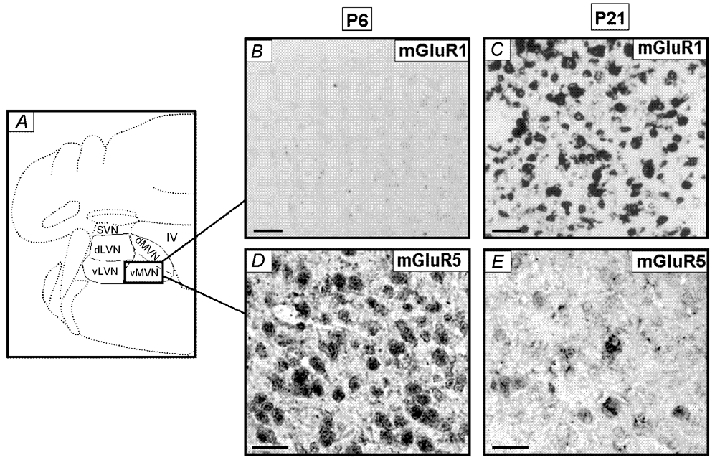
Figure 6. Distribution of hybridization signals for mGluR1α and mGluR5 mRNA in the rat MVN during postnatal development.

A, schematic representation of the various regions of the VN on adult coronal section of the brainstem. vMVN, ventral part of the MVN; dMVN, dorsal part of the MVN; dLVN, dorsal part of the LVN; vLVN, ventral part of the LVN; SVN, superior VN; IV, fourth ventricle. B-E, brainstem sections at different postnatal (P) days were hybridized using specific oligoprobe for mGluR1 (B and C) and mGluR5 (D and E). Scale bars, 25 µm.
