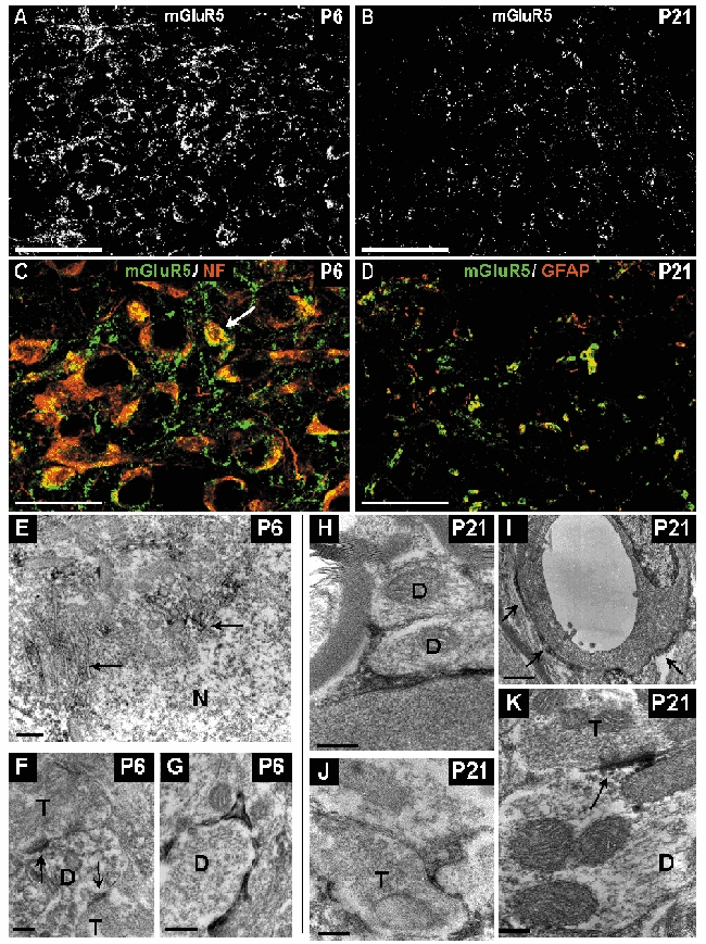
Figure 9. Distribution of mGluR5 immunoreactivity in the developing vMVN visualized by confocal microscopy immunofluorescence and electron microscopy immunoperoxidase staining.

A, area of vMVN exhibiting a very high density of staining at P6. Note also the numerous mGluR5 immunoreactive neurons. B, sparse immunoreactive puncta at P21. C, colocalization of anti-mGluR5 (green) and anti-NF (red) antibodies in neurons at P6. All large neurons express mGluR5 but rare small neurons (arrow) are also labelled. D, double labelling with GFAP (red) and mGluR5 (green) antibodies shows a colocalization in the glial cells of the vMVN. Scale bars, 50 µm (A and B) and 25 µm (C and D). E, thin small accumulations of immunoreaction product associated with rough endoplasmic reticulum and Golgi apparatus (arrows) in the cytoplasm of the neuron at P6. F, mGluR5-labelled postsynaptic densities (arrows) in a dendritic profile opposing two axonal terminals (T). G, immunoreactive glial processes ensheathing an unlabelled dendrite (D) at P6. H, immunoreactive glial processes ensheathing unlabelled dendrites at P21. I, immunoreactive glial processes (arrows) around the wall of a capillary. J, labelled glial processes enwrapping an unlabelled axonal terminal. K, strongly immunoreactive postsynaptic density opposed to unlabelled axonal terminal at P21. Scale bars, 500 nm (A and E) and 200 nm (B, C, D, G and F).
