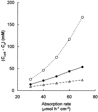
Figure 5. Concentration differences across subjunctional lateral membranes of rat enterocytes during absorption of glucose.

At glucose absorption rates greater than 30 µmol h−1 cm−2, the predicted concentration differences across subjunctional lateral membranes of rat jejunal enterocytes increase with absorption rate. The three curves show the relations when the vascular response to glucose loading is half normal (^), normal (•) and twice normal (▵). When the normal increase in Qb and PS is halved, the concentration in the subjunctional intercellular fluid exceeds 160 mm when the glucose flux is 70 µmol h−1 cm−2 so that to maintain the flux, the apical cytosolic concentration exceeds 300 mm (see Figs 3 and 4). Such high intracellular concentrations may be expected to diminish the apical influx on SGLT-1 and under these conditions of low blood flow the villus microvascular system limits the rate at which epithelial transport can be sustained.
