
Figure 4. In the near absence of SNARE-dependent neurotransmitter release, BDNF induces spine formation, while increasing the proportion of thin spines (type-III).
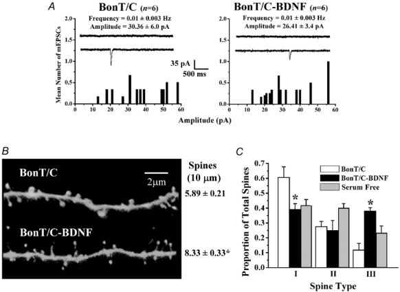
A, probability distributions of the mean amplitudes of AMPA-mediated mEPSCs in slice cultures treated with botulinum neurotoxin C (BoNT/C) (left) and BoNT/C-BDNF (right). B, representative dendritic segments of CA1 pyramidal neurones in BoNT/C (top) and BoNT/C-BDNF (bottom) slice cultures. C, histogram plots of the proportion of each morphologically distinct spine type in slice cultures treated with BoNT/C and BoNT/C-BDNF. Data from serum-free controls (from Fig. 2) are presented here to facilitate visual comparisons.