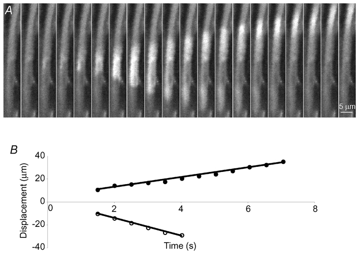
Figure 6. Spontaneous Ca2+ waves in a smooth muscle cell.

A, an example of a cell in which Ca2+ waves arose spontaneously and propagated bidirectionally. The locations of the wave fronts are plotted in B. In this cell the average speeds of the waves were 4.4 µm s−1 (•) and 7.4 µm s−1 (^). A and B have the same time scale.
