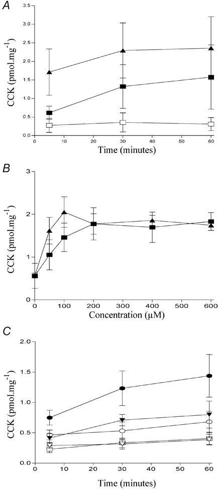
Figure 5. The effect of other modifications on fatty-acid induced CCK secretion.

Comparison of the ability of dodecanoic acid (▪) and perfluorododecanoic acid (▴) to induce CCK secretion in STC-1 cells exposed for up to 60 min (A) with each fatty acid or to the ethanol vehicle control (□). The potency of each fatty acid was determined by exposing STC-1 cells to various concentrations (0–600 μm) for 30 min (B). The effect of shorter chain perfluoro fatty acids on CCK secretion in STC-1 cells is shown in C. STC-1 cells were exposed for up to 60 min with heptanoic acid (▿); octanoic acid (○); perfluoro heptanoic acid (▾); perfluoro octanoic acid (•) or to an ethanol vehicle control (□). Determination of CCK secretion is described in Fig. 2. Each data point represents the mean ±s.e.m. of four experiments.
