
Figure 9. The effect of various chemical modifications on fatty acid-induced Ca2+ mobilisation.
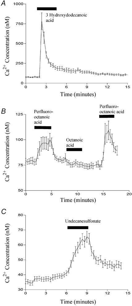
The effect of 500 μm 3-hydroxydodecanoic acid (A), 500 μm perfluoro-fatty acids (B) and 500 μm 1-undecanesulfonic acid (C) on [Ca2+]i in STC-1 cells. Cells were exposed to each fatty acid for 3 min. In B, cells were exposed to perfluoro-octanoic acid (first bar), octanoic acid (second bar) and perfluoro-octanoic acid a second time (third bar). Fluorescent images were acquired and processed as described in Fig. 8. The number of cells that make up each mean is 12 (A), 13 (B) and 14 (C) and these figures are representative of three other experiments.