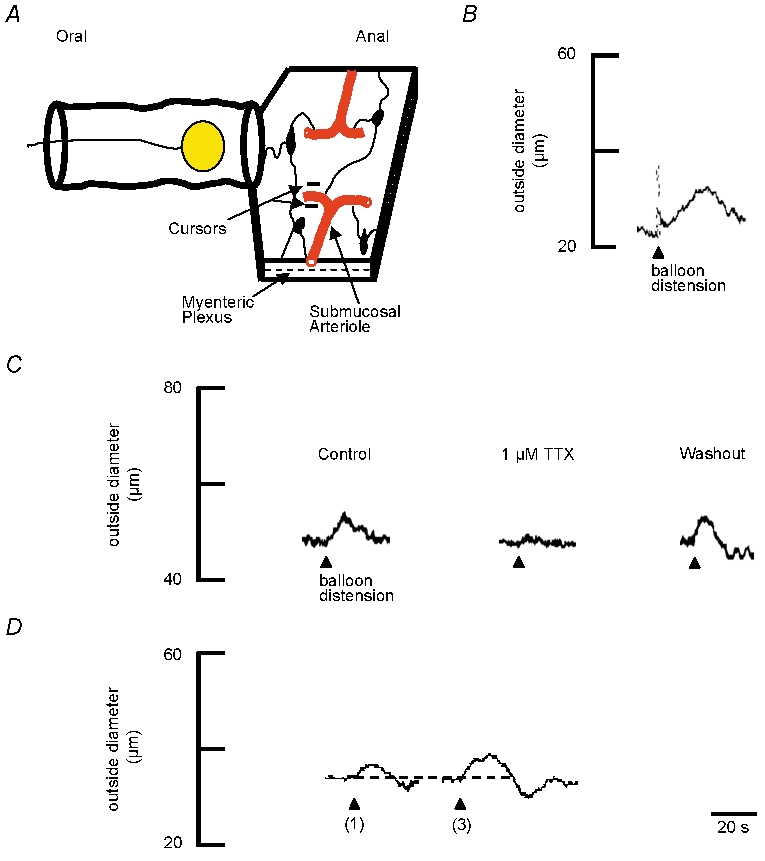
Figure 1. Vasodilatation evoked by luminal distensions.

A, schematic representation of ileal preparation. The mucosa was removed from the aborad segment to expose submucosal arterioles and submucosal ganglia (dark elliptical shapes). Changes in outside diameter of the arteriole were monitored at a single site (cursors) by videomicroscopy. Arterioles were preconstricted 80–95 % of maximum with the prostaglandin analogue PGF2α (400 nm; present in the superfusate for the duration of the experiment). The intestine was distended with a balloon placed within the lumen. The distance from the centre of the balloon to the recording site on the arteriole was 1–2 cm. B, representative trace showing that one distension (arrowhead) evokes dilatation of the arteriole. The resting outside diameter was 44 μm. C, representative traces showing TTX (1 μm) blocks the distension-induced vasodilatation compared to control. The resting outside diameter was 48 μm. A 15 min washout period restores the vasodilatation evoked by distensions. D, representative traces showing that increasing the number of balloon distensions from one distension to three distensions (≈1 Hz) increases the size of dilatations. Dotted line indicates the baseline prior to distension. The resting outside diameter was 46 μm.
