
Figure 3. Effect of mucosa on distension-induced vasodilatations.
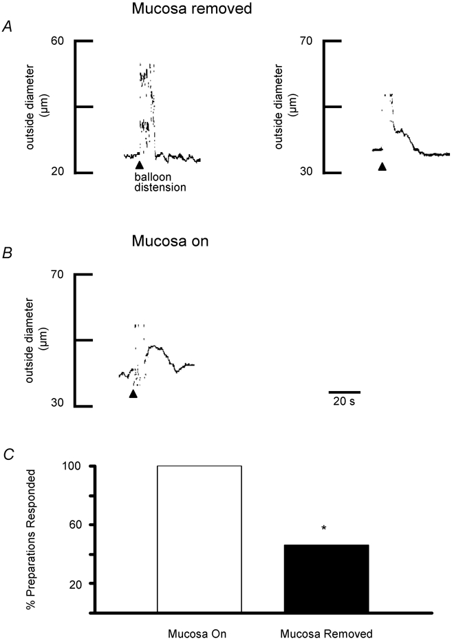
A, representative traces of preconstricted arterioles from two separate preparations illustrating that varying responses were obtained when the mucosa was removed from the entire preparation. In the preparation on the left, balloon distension (arrowhead, 3 distensions) failed to elicit a dilatation whereas a vasodilatation was elicited in the preparation shown on the right. Resting outside diameters were 50 μm (left) and 57 μm (right). B, when the mucosa was not removed from the stimulating site, balloon distensions consistently dilated arterioles, as shown in this representative trace (arrowhead, 3 distensions). Resting outside diameter was 60 μm. C, summary of the percentage of preparations in which balloon distension evoked a dilatation with the mucosa intact (n = 12 preparations) compared to those where it had been removed (n = 13 preparations). *P = 0.005