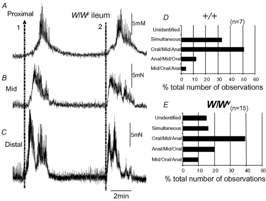
Figure 5. Comparison of the direction of propagation of MMCs recorded from the ileum of +/+ and W/Wv mice.

A, B and C, two MMC contractions, one that propagates from the anal to the oral region of the ileum (orally migrating, represented by arrow 1), while another (represented by the dotted line 2) appears to have occurred simultaneously in the proximal, mid and distal regions. D, the proportion of MMCs in +/+ mice where the direction of propagation was noted. E, shows the same graphical representation of MMCs in W/Wv mice and the direction of MMC propagation. In both types of mice, the predominant direction of propagation was from the proximal to mid to distal ileum.
