
Figure 3. Identification of muscle fibre membrane lesions by the presence of intracellular procion orange.
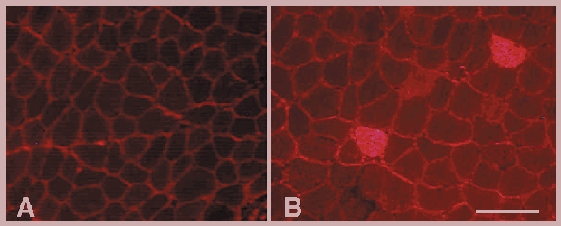
A, cross-section of a control soleus muscle that was incubated with the extracellular marker dye procion orange. In healthy control muscle the procion orange is located in the extracellular space that outlines each muscle fibre. B, cross-section of a soleus muscle from a C57 mouse that experienced 24 h of muscle reloading after 10 days of unloading. Two fibres are brightly fluorescent because of the entry of procion orange through membrane lesions. Note also that the general level of intracellular fluorescence in muscle fibres in reloaded soleus (B) is higher than in control fibres (A). Bar = 100 μm.