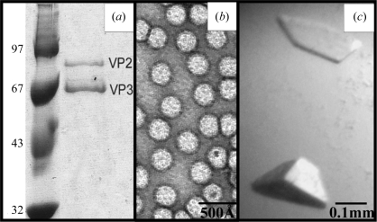
Figure 1.
Purification and crystallization of AAV7 viral capsids. (a) SDS–PAGE gel showing the AAV7 viral proteins VP2 and VP3 (molecular weights 73 and 62 kDa, respectively) assembled into capsids in the baculovirus expression system in the right lane. VP1 was not present in the capsids. The left lane shows the position of molecular-weight standards (kDa). (b) Transmission electron micrograph of intact AAV7 capsids stained with 2% uranyl acetate. (c) Optical photograph of an AAV7 capsid crystal.

