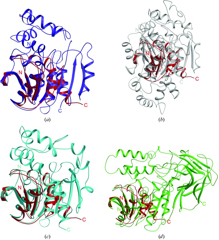
Figure 2.
Superimposition of the structure of TTHA1544 (red) with those of four other proteins: (a) A. radiobacter AD1 epoxide hydrolase (PDB code 1ehy, blue), (b) murine liver epoxide hydrolase (PDB code 1cr6, grey), (c) Burkholderia sp. FA1 fluoroacetate dehalogenase (PDB code 1y37, cyan) and (d) Rhodococcus sp. MB1 cocaine esterase (PDB code 1ju3, light green), shown in a ribbon presentation. These figures were prepared with the programs MOLSCRIPT (Kraulis, 1991 ▶) and RASTER3D (Merritt & Bacon, 1997 ▶).

