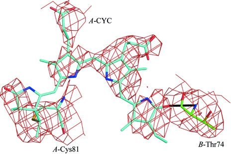
Figure 2.
A 2F o − F c, σA-weighted electron-density map of a model perturbed by shifts in each atom of up to 0.2 Å with the chromophore omitted. This map should show an unbiased view of the chromophore. The map is contoured at 1.2σ around Cys84 of the α subunit and the covalently attached phycocyanobilin chromophore (in blue) and the Thr74 of chain B (in green), with the hydrogen bond indicated between the chromophore and the backbone N atom of Thr74.

