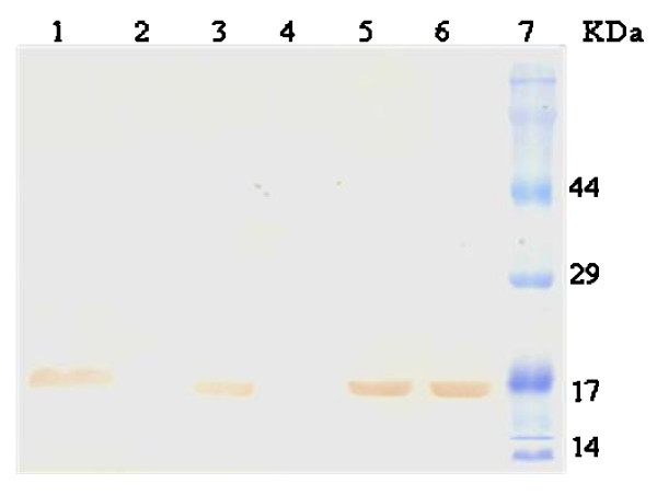
Figure 4.
Western blot of homogeneous rhG-CSF. A single band was observed for rhG-CSF transferred to a nitrocellulose membrane and detected using a polyclonal antiserum. Lane 1: crude extract of host cells transformed with pET 23a(+)::hG-CSF; lane 2: crude extract of host cells transformed with pET 23a(+) (control); lane 3: insoluble fractions of host cells transformed with pET 23a(+)::hG-CSF; lane 4: insoluble fractions of pET 23a(+); lane 5: homogeneous hG-CSF; lane 6: reference standard; and lane 7: protein molecular mass markers (Cruz Marker™).

