
Figure 1.
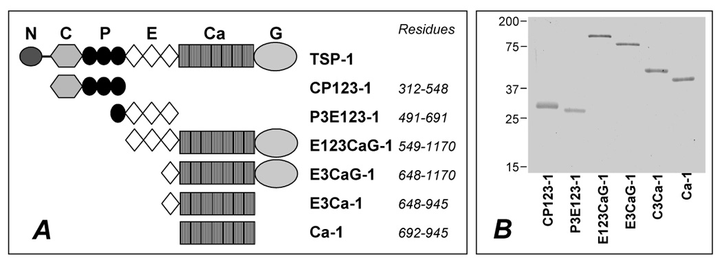
A) Schematic representations of the human TSP-1 monomer and the recombinant fragments used in this study. Abbreviation for the structural domains are: N, amino-terminal domain; C, procollagen like domain; P, properdin like, type I repeat; E, EGF-like, type II repeat; Ca, calcium-binding, type III repeats; G, globular carboxy-terminal domain. Residues are related to the initiating methionine. B) Purified recombinant proteins were resolved by SDS-PAGE and stained with Coomassie blue. Molecular weight markers are indicated in kDa. The type III repeats-containing domains migrated more slowly than predicted by their molecular weight, as reported (Anilkumar, Annis, Mosher & Adams, 2002; Hannah, Misenheimer, Annis & Mosher, 2003).