
Figure 3.
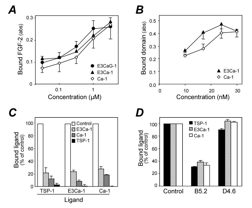
Localization of the FGF-binding site in the type III repeats of TSP-1. A) Binding of labeled FGF-2 to plastic coated with increasing concentrations of TSP-1 fragments. Data are the amount of bound FGF-2, as absorbance, mean and SD of triplicates. B) Binding of labeled E3Ca-1 or Ca-1, at the indicated concentrations, to immobilized FGF-2. C) Competition assay: binding of labeled TSP-1, E3Ca-1 and Ca-1 to immobilized FGF-2 in the presence of 2.5 µM unlabeled E3Ca-1, Ca-1, or TSP-1 used as competitors (see legend). Data are the percentage of control binding, in the absence of competitors. D) Effect of the monoclonal anti-TSP-1 antibodies B5.2 and D4.6 (50 µg/ml) on the binding of TSP-1, E3Ca-1, or Ca-1 to immobilized FGF-2. Binding is the percentage of control, in the absence of antibodies. Experiments were done in calcium-free buffers.