
Figure 6.
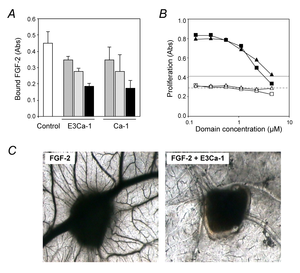
Antiangiogenic activity of the TSP-1-type III repeats. A) Binding of FGF-2 to endothelial cells. BAEC were incubated with labeled FGF-2 in the absence (control, white column) or presence of the indicated recombinant fragment (7.5 µM grey columns, 15 µM striped columns, 30 µM black columns). The amount of cell-bound FGF-2 is expressed as absorbance. B) Endothelial cell proliferation. BAEC were exposed to the indicated concentration of E3Ca-1 (triangles), Ca-1 (squares), or TSP-1 (25 µg/ml, lines) with (black symbols, solid line) or without (white symbols, dashed line) 5 ng/ml FGF-2 and incubated for 3 days. Proliferation is expressed as absorbance. C) FGF-2-induced angiogenesis in the CAM assay. FGF-2 was administered in the absence or presence of E3Ca-1 on day 8 and pictures were taken 4 days later (n=10). Original magnification: 50 x.