
Figure 1.
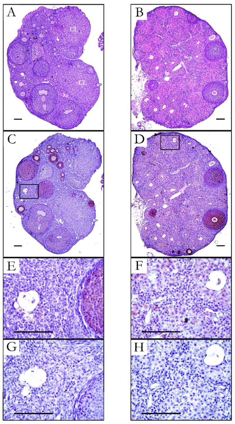
Photomicrographs of ovaries from Siberian hamsters held in either long days (LD; left panels) or short days (SD; right panels) from conception to 10 wk of age. (A and B) Hematoxylin and eosin staining. Corpora lutea (CL) were seen only in LD ovaries. (C and D) Immunhistochemistry for anti-Müllerian hormone (AMH) showing the most intense staining in granulosa cells in primary and secondary follicles. (E and F) Higher magnification of boxed regions in C and D, showing oocyte remnants and cells that surround them. Granulosa cells surrounding atretic oocytes in SD (F) have hypertrophied (luteinized) and stain positive for AMH, in contrast to comparable cells in LD ovaries (E). (G and H) Sections adjacent to those shown in E and F for which the primary antibody against AMH was pre-incubated with the blocking peptide – refer to methods for details. Bars = 100 μm.