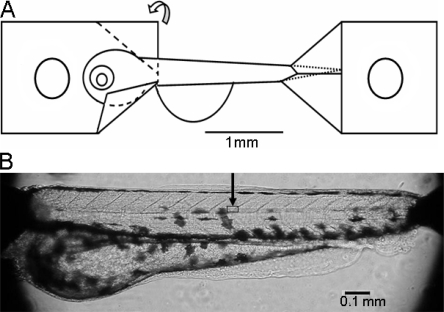
Figure 1.
Zebrafish larvae preparation. (A) Schematic illustration of preparation attachment using aluminum foil clips at both ends of the larvae. The aluminum foil is folded along the dashed lines as shown on the left clip. A small hole is made in each clip for mounting. (B) Photograph of the preparation mounted on the microscope stage. The square area under the arrow indicates the approximate location of the area examined with higher magnification light microscopy in Fig. 2 A.

