Figure 3.
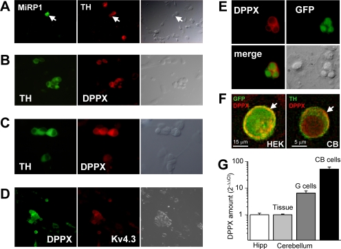
MIRP1 and DPPX expression in rabbit CB chemoreceptor cells. (A) The presence of MiRP1 protein in cultured CB chemoreceptor cells was explored by double labeling with anti-TH and anti-MiRP1 antibody. MiRP1-positive cells were scant and of those very few were chemoreceptor cells (as the one marked with the arrow). The corresponding transmitted light image is also shown for each field. (B) Immunofluorescence labeling of DPPX shows the expression of DPPX in every TH-positive cell (and also in some TH-negative cells). Anti-DPPX antibody provided by E. Wettwer. (C) Same results as in B were obtained with anti-DPPX antibody from Santa Cruz Biotechnologies. (D) Double labeling of CB chemoreceptor cells with anti-Kv4.3 and anti-DPPX antibodies shows an almost perfect coexpression of these two proteins, as expected if they associate in heteromultimeric channels. (E) Specificity of the DPPX antibodies was explored in HEK cells transfected with GFP+DPPX. (F) Deconvolved images showing a preferential localization of DPPX (arrows) in the surface of a Kv4.3+DPPX-transfected HEK cell (left) and a chemoreceptor cell (right). Red fluorescence corresponds to DPPX labeling, and green fluorescence corresponds to GFP (left) or TH (right) labeling. Anti-DPPX antibody from E. Wettwer was used in E and F, but the same results were obtained with the other antibody tested. (G) Real-time PCR showing the relative abundance of DPPX mRNA in CB chemoreceptor cells in primary culture. Normalized amount of DPPX mRNA in rabbit hippocampus (Hipp) was used as calibrator, and DPPX mRNA abundance in whole cerebellum (Tissue) and in cerebellar granule cells (G cells) were determined for comparisons. For details of the 2−ΔΔCt relative quantification method see Materials and methods section. Each bar is the mean ± SEM of four to seven individual determinations.

