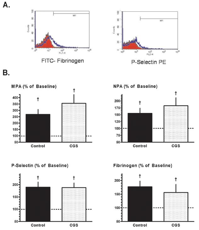
Figure 2.

Flow cytometry: Protocol 1. (A) Representative histograms illustrating the increase in platelet-fibrinogen binding (left) and platelet surface P-selectin expression (right) in paired canine blood samples obtained at 2 hours after the onset of recurrent thrombosis (blue profiles) versus baseline (red profiles). (B) Monocyte-platelet aggregates (MPA), neutrophil-platelet aggregates (NPA), platelet surface P-selectin expression and platelet-fibrinogen binding, measured at 2 hours after the onset of recurrent thrombosis and expressed as a % of baseline values, in Control and CGS 21680-treated dogs. Data for control group reported previously in [8]. †p<0.05 versus baseline; no significant differences between groups.
