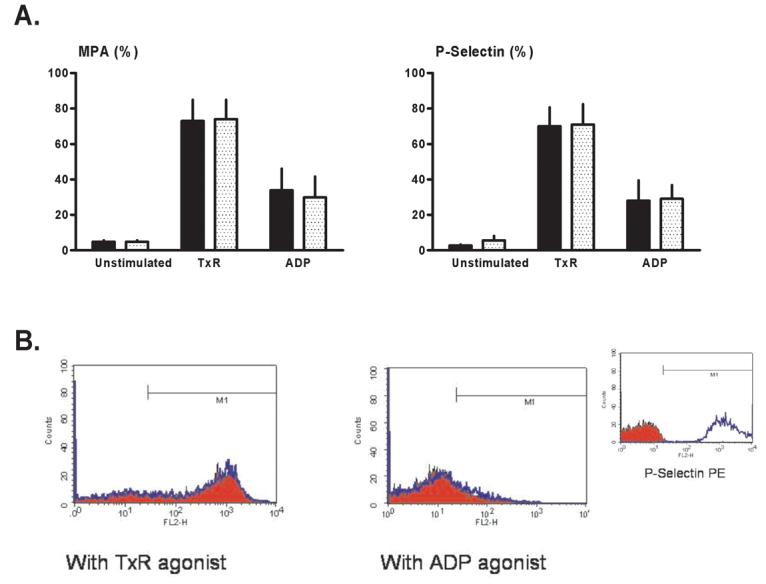
Figure 4.

Flow cytometry: Protocol 2 (canine blood samples). Monocyte-platelet aggregates (MPA) and platelet surface P-selectin expression were quantified in unstimulated (no agonist, quiescent) blood aliquots, aliquots stimulated with a stable thromboxane receptor agonist (TxR), and aliquots stimulated with ADP. (A) Mean values for samples pre-treated with CGS 21680 (stippled bars) versus paired vehicle-controls (solid bars). (B) Representative histograms showing platelet surface P-selectin expression in CGS-treated blood aliquots (red profiles) versus vehicle-controls (blue profiles). (Insert) Positive and negative controls: histograms for isotype-negative (red) and PMA-positive (phorbol myristate acetate: blue) controls for P-selectin in canine blood.
