
Figure 1.
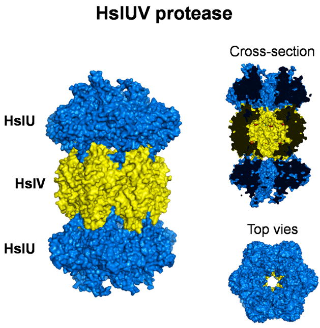
Structures of the bacterial ATP-dependent protease HslUV (PDB 1G3I). The protease subunits HlsV are shown in yellow, the ATPase subunits HslU are shown in blue. A side-on cross section reveals the active site of proteolysis (red dots) in the catalytic chamber and the degradation channel that connects the active site to the exterior of the protease. End-on view shows the sixfold axis of symmetry. Structures were produced by PyMOL.