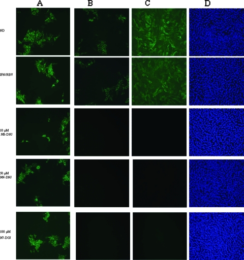
FIG. 4.
IF analysis of naïve MDBK cells incubated with supernatants from treated BVDV-infected cells (set 2) at P10 (A) and P22 (B) and of long-term-treated BVDV-infected MDBK cells (set 2) at P22 (C). The cells were fixed and probed with a monoclonal antibody against the BVDV NS2 and NS3 proteins, followed by incubation with an anti-mouse FITC-conjugated secondary antibody (green). Cell nuclei were stained with DAPI (D). ND, no drug.

