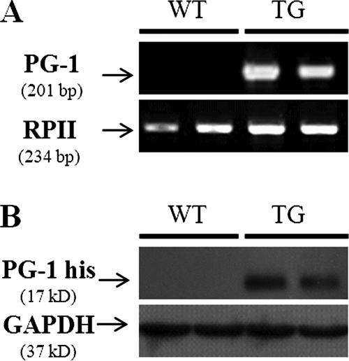
FIG. 2.
Expression of PG-1 in the lungs of mice. (A) Representative gel image of RT-PCR results. Total RNA was isolated from the lungs of WT and transgenic (TG) mice, and RT-PCR was conducted with PG-1-specific primers. The PCR products were resolved on a 1% agarose gel and stained with ethidium bromide for visualization. No PG-1 transcript was detected in WT mouse lung tissue, while a clear 201-bp band could be visualized in transgenic mouse lunf tissue samples. Amplification of RNA polymerase II (RPII) was used as a loading control. (B) Representative Western blot showing the expression of PG-1-His in transgenic mouse lung tissue. Proteins were extracted from the lungs of WT and transgenic mice, separated by sodium dodecyl sulfate-polyacrylamide gel electrophoresis, and immunoblotted with anti-His tag antibody. The housekeeping protein GAPDH was used as the loading control.

