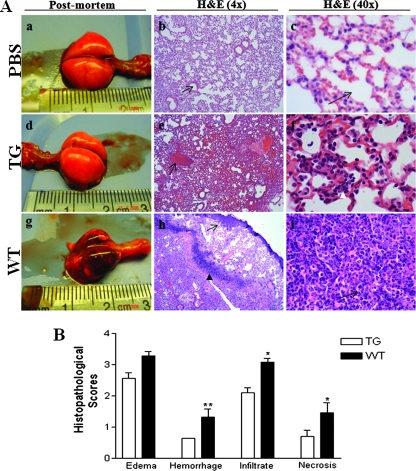
FIG. 4.
Comparison of gross and microscopic pulmonary injury after inoculation of A. suis. (A) Representative photographs of the gross appearance of whole lungs of mice inoculated with PBS (a), PG-1 transgenic (TG) mice inoculated with A. suis (d), and the WT littermate mice inoculated with A. suis (g) at postmortem examination. Tissue sections from mice inoculated with PBS were normal, with no evidence of edema, hemorrhage, leukocytic infiltrates, or necrosis; and the alveolar spaces were clear (b and c; arrows). Tissue sections from transgenic mice inoculated with A. suis showed focal congestion (e; arrow) and moderate neutrophilic infiltrates (f; white arrowhead). Tissue sections from WT mice inoculated with A. suis showed marked congestion (h; arrow), intact bacteria (h; black arrowhead), and marked neutrophilic and macrophage infiltrates within the alveolar spaces (i; arrow). H&E, hematoxylin-eosin. (B) Overall histopathological score (Table 1) for lungs from transgenic mice (open bars) and WT mice (solid bars) inoculated with A. suis. Data represent the means ± SEMs (PBS-inoculated mice, n = 6; transgenic mice, n = 33; WT mice, n = 24). **, P < 0.01; *, P < 0.05.

