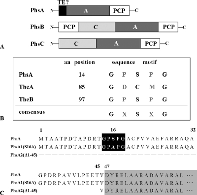
FIG. 3.
(A) Domain organization of the peptide synthetases PhsA, PhsB, and PhsC. (B) Localization of the TE motif GXSXG in PhsA, TheA, and TheB. In TheA, the active serine site is replaced by a cysteine residue. (C) Schematic representation of the primary sequence of the native PhsA protein as well as the mutated PhsA variants PhsA1(S16A) and PhsA2(Δ1-45). The TE motifs in PhsA and PhsA1(S16A) in which the conserved serine residues were changed to alanines are highlighted by a black box. The beginning of the adenylation domain of PhsA at aa 47 is marked by a gray box.

