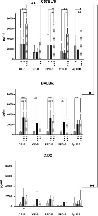
FIG. 2.
Mycobacterium-specific IFN-γ secretion by splenocytes from M. paratuberculosis-infected C57BL/6, BALB/c, and C.D2 mice. IFN-γ production was measured in spleen cell culture supernatants from C57BL/6, BALB/c, and C.D2 mice before and 4 weeks (gray bars), 8 weeks (black bars), and 12 weeks (hatched bars) after infection with M. paratuberculosis S-23. The cells were stimulated for 72 h with CF-P or CF-B (10 μg/ml), PPD-P or PPD-B (10 μg/ml), or recombinant Ag85B (5 μg/ml) from M. paratuberculosis. Shown are means plus standard deviations of three or four mice tested individually. Statistical analyses were performed using two-way ANOVA with Bonferroni posttests. Comparison to naïve mice: *, P < 0.05; **, P < 0.01; ***, P < 0.001. Comparison to previous time point: ○, P < 0.05; ○○, P < 0.01; ○○○, P < 0.001. Comparison between M. paratuberculosis Ag and M. bovis Ag: ⧫, P < 0.05. Ag85B-specific responses compared between mouse strains: •, P < 0.05; ••, P < 0.01.

