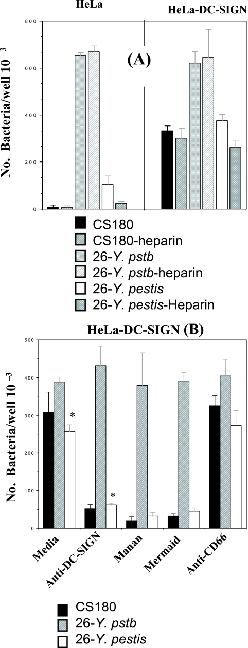
FIG. 4.
DC-SIGN-mediated phagocytosis of Y. pestis is inhibited by heparin, anti-DC-SIGN antibody, mannan, and DC-SIGN-like Mer- maid. (A) E. coli K-12 CS180, Y. pseudotuberculosis serotype O:1a, and 26-Y. pestis were incubated with HeLa and HeLa-DC-SIGN cells for 2.5 h in the presence or absence of heparin (30 μg/ml). (B) 26-Y. pestis was incubated with HeLa-DC-SIGN cells for 2.5 h in the presence or absence of anti-CD66 (5 μg/ml) and DC-SIGN (5 μg/ml), mannan (500 μg/ml), and DC-SIGN-like protein (His-Mermaid; 10 μg/ml). All reagents were added to the media for 20 min before the addition of bacteria. Heparin (30 μg/ml) was added to every sample in order to eliminate the non-DC-SIGN-specific interaction. The concentration of each reagent used in the experiment was based on previously published data (32, 61, 62). The phagocytosis rate of Y. pestis was evaluated by the recovery of bacteria from gentamicin protection. E. coli K-12 CS180 and Y. pseudotuberculosis serotype O:1a were control strains. In addition, only half the number of Y. pseudotuberculosis cells were added to the HeLa-DC-SIGN sample in order to better compare the effects of these reagents among the three strains. (A) Y. pseudotuberculosis promotes effective internalization into HeLa cells. *, P < 0.001, based on Student's t test comparing the interaction of HeLa-DC-SIGN cells with 26-Y. pestis in the presence of anti-DC-SIGN antibody to the interaction without the presence of antibody. n was nine samples for each test. The error bars indicate standard errors.

