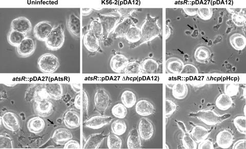
FIG. 7.
Phase-contrast microscopy of infected ANA-1 macrophages. The infections were performed at an MOI of 50:1 for 4 h with parental and mutant strains containing pDA12, pAtsR, or pHcp as indicated in parentheses. atsR indicates DFA21; atsR Δhcp indicates DFA28. Black arrows indicate vacuole-containing protrusions. WT, wild type.

