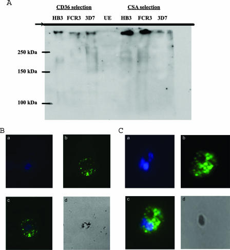
FIG. 7.
(A) Western blot of PEs using the anti-GST-CIDR-Δ106 polyclonal antibodies. Mature-stage parasites were extracted and lysed. Proteins were separated by sodium dodecyl sulfate-polyacrylamide gel electrophoresis and transferred onto nitrocellulose membranes. The top of the separating gel is indicated. Murine polyclonal antisera against GST-CIDR-Δ106 were used to detect the expression of PfEMP-1 at a dilution of 1:200, followed by secondary antibodies and enhanced chemiluminescence. PfEMP-1 can be detected on both CD36-adherent and CSA-adherent PEs of strains 3D7, HB3, and FCR3, while no signal can be detected in uninfected erythrocytes. (B and C) Surface labeling of CD36- and CSA-adherent PEs of strain 3D7 with murine polyclonal antisera raised against GST-CIDR-f:106-166. a, staining of parasite nuclei using DAPI; b, surface labeling of fluorescein isothiocyanate; c, merged image; d, phase image.

