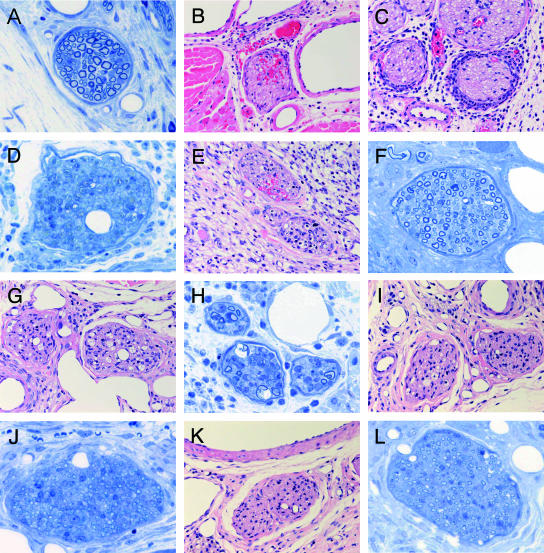
FIG. 3.
Sequential analysis of footpad histopathology after mycolactone injection (100 μg). (A) Nerve of control mouse (only ethanol and 7H9 broth injected) showed no significant change. (B to D) On day 7, nerves of mycolactone-injected mice showed intraneural hemorrhage (B) and massive neutrophilic infiltration to the perineurium (C). (D) Epon section showing moderate loss of nerve fibers and myelin. (E and F) Nerves on day 14 showed intraneural inflammatory cell infiltration and hemorrhage. (G) Nerves on day 21. Mild infiltration inflammatory cell infiltration and vacuolar change were observed. (H) Vacuolar change of Schwann cells induced by mycolactone injection on day 21 was similar to M. ulcerans inoculation. (I) In the nerves on day 28, histological damage remained. (J) Thin myelins indicating remyelination were observed on day 28. (K) In the nerves on day 42, when sensory disturbance lasted, histological damage remained. (L) Mild fibrosis was observed on day 42. (A, D, F, H, J, and L [Epon, 1-μm sections, toluidine blue staining], magnification, ×340; B, C, E, G, I, and K [paraffin sections, H&E staining], magnification, ×157).

