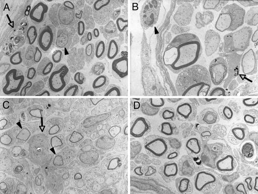
FIG. 4.
Ultrastructure of nerves after mycolactone injection. (A) Intraneural infiltration of lymphocytes (arrowhead) and macrophage (arrow) containing myelin debris on day 14 after mycolactone injection. (B) Vacuolar change of myelin (*) and macrophage (arrow) on day 21. (C) Thin myelins (arrows) indicate remyelination on day 28. D. Mild fibrosis observed on day 42. (Epon-embedded ultrathin sections stained by uranium and lead; magnification, ×1,960).

