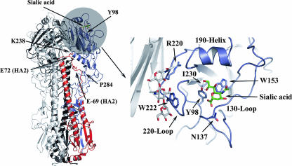
FIG. 1.
Left panel, ribbon diagram showing the HA trimer. One of the three monomers is colored to show the HA1 subunit in blue and the HA2 subunit in red. The region encompassing the receptor binding domain is shaded, and the location of bound sialic acid is indicated in green. The location of HA1 Tyr-98 at the base of the binding pocket is also indicated, as are some of the positions identified in reversion mutants that may influence trimer stability. Right panel, enlarged view of the receptor binding region. The binding site is a shallow depression bordered by a short α-helix (the 190 helix) at the membrane-distal edge, the 130 loop at the front of the site, and the 220 loop at the left side. Conserved residues Y98, W153, H183, and Y195 form the base of the site, and the positions of Y98 and W153 are indicated. Sialic acid is shown in green, and the location of residues identified in reversion mutants are indicated (as they appear in WT HA). The sialic acid binding pocket is located in the monomer colored in blue, but residues from the adjacent monomer (shown in gray) can influence binding, particularly if carbohydrate attachments are involved. This is illustrated by the location of the carbohydrate chain originating from HA1 Asn-165 of the neighboring monomer, shown here in gray and red at the left side of the binding site.

