FIG. 2.
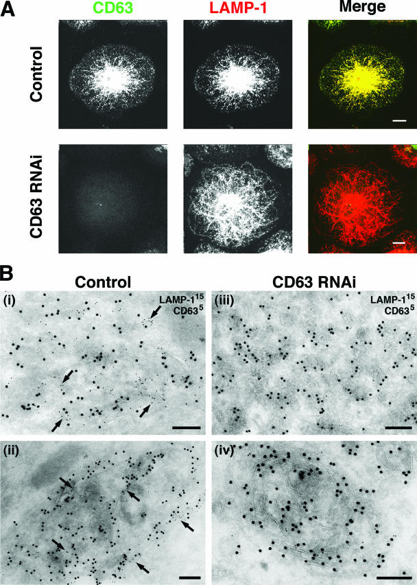
Distribution of lysosomal markers in CD63 knockdown MDM. MDM were nucleofected with scrambled or CD63-specific siRNAs and returned to culture for 2 to 12 days. (A) Cells were fixed 6 days after nucleofection, permeabilized, and stained with MAbs against CD63, followed by anti-mouse IgG2b Alexa Fluor 488, and against LAMP-1, followed by anti-mouse IgG1 Alexa Fluor 594. Images show projections of 22 and 23 confocal sections (0.5 μm thick) for the control and CD63 RNAi samples, respectively. Scale bars = 10 μm. Observations similar to those above were made 2 and 4 days after nucleofection. (B) EM immunolabeling of control (panels i and ii) or CD63 knockdown MDM (panels iii and iv) 12 days after nucleofection. Ultrathin cryosections were double labeled with MAbs against LAMP-1/PAG (15 nm) and CD63/PAG (5 nm). Arrows indicate CD63 staining. Scale bars = 200 nm.

