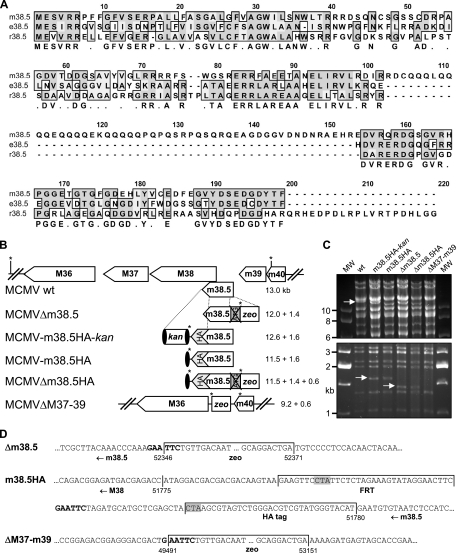
FIG. 1.
Evolutionary conservation and mutagenesis of MCMV m38.5. (A) Clustal W amino acid sequence alignment of MCMV Smith strain protein m38.5 (corresponding to nt 51780 to 52367; GenBank accession no. NC_004065), rat CMV English isolate protein e38.5 (corresponding to GenBank accession no. EU267790), and rat CMV Maastricht isolate protein r38.5 (corresponding to nt 35738 to 36235; GenBank accession no. NC_002512). Sequences were aligned using a Blossum matrix. Identical and similar amino acids are boxed and shaded in dark and light gray, respectively. The consensus sequence is shown below. (B) Arrangement of the M36 through m40 ORFs of wt MCMV and construction of m38.5 mutant viruses by BAC mutagenesis. The m38.5 gene was knocked out by deleting the ATG start codon and inserting a bacterial zeo gene, resulting in the MCMVΔm38.5 deletion mutant. A HA epitope tag sequence was attached to the 3′ end of the m38.5 gene by using a kan cassette flanked by FRT sites (black ovals) as a selectable marker. The kan cassette was removed by Flp recombinase. A second m38.5 knockout mutant, MCMVΔm38.5HA, was constructed on the basis of MCMV-m38.5HA. A larger deletion comprising M37 through m39 ORFs was generated by inserting a zeo cassette. EcoRI restriction sites are indicated by asterisks, and the sizes of the expected EcoRI fragments are listed. (C) EcoRI restriction patterns of wt and mutant MCMV BACs separated by agarose gel electrophoresis. The 13-kb fragment of wt MCMV and the 1.4- and 1.6-kb fragments of the mutant viruses are indicated by arrows. MW, molecular size standard; m38.5HA-kan, MCMV-m38.5HA-kan; m38.5HA, MCMV-m38.5HA; Δm38.5, MCMVΔm38.5; Δm38.5HA, MCMVΔm38.5HA; ΔM37-m39, MCMVΔM37-m39. (D) The mutated sites of all five mutant viruses were sequenced. Three sequences are shown, and the remaining sequences contained related or combined mutations. Numbers indicate nucleotide positions within the MCMV genome, and the inserted sequences are labeled. The FRT site is flanked by short linker sequences. EcoRI restriction sites are shown in bold. The stop codon of the m38.5 ORF (at the end of the HA tag sequence) and a stop codon within the FRT site that terminates the M38 ORF are shaded in gray.

