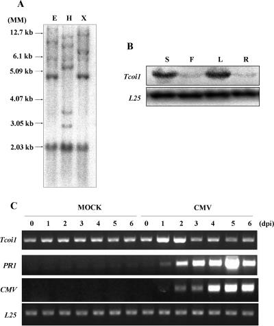
FIG. 2.
Southern blot analysis and comparison of Tcoi1 expression patterns in various organs and in tissues from CMV-Kor- and mock-inoculated plants. (A) Tobacco genomic DNA was digested with EcoRI (E), HindIII (H), or XbaI (X), and digestion products were separated on 0.8% agarose gel. After being transferred onto a Nytran Plus membrane, the blot was hybridized with a 32P-labeled full-length Tcoi1 cDNA probe under conditions of medium stringency. Autoradiograms were visualized with a Fuji BAS 2500 phosphorimager. DNA size standards (MM) are shown at the left. (B) Accumulation of Tcoi1 transcripts in different organs. Lanes: S, stem; F, flower; L, leaf; and R, root. Tcoi1 transcripts were monitored by Northern blot analysis using the Tcoi1-specific 3′ untranslated region as a probe. The transcript level corresponding to ribosomal protein L25 was included as an internal standard for RNA quantity evaluation. (C) RT-PCR analysis of Tcoi1 genes upon CMV-Kor inoculation. Total RNA was extracted from leaf tissues at 0, 1, 2, 3, 4, 5, and 6 dpi. Tissue was isolated from the leaves of either CMV- or mock-inoculated plants. The conserved 3′ ends of CMV RNAs and PR-1 were detected as a positive control for CMV inoculation. As an internal standard for cDNA quantity evaluation, the level of L25 was monitored.

