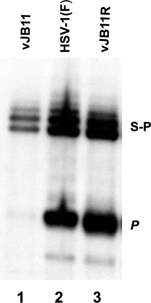
FIG. 6.
Southern blot of viral DNA digested with BamHI. Approximately 2 × 10⁶ CV1 cells were infected with the indicated viruses, and viral DNA was extracted, digested with BamHI, transferred to a nylon membrane (0.45 μm), and hybridized with radiolabeled BamHI P fragment of HSV-1(F) DNA. Bound DNA was visualized by autoradiography. The positions of the S-P fragment corresponding to junctions of the S and L component in concatameric DNA and the P fragment indicative of genomic end fragments are indicated.

